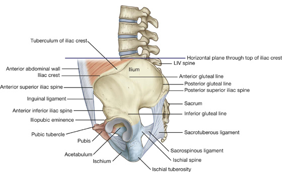
Each pelvic bone is formed by three bones (ilium, ischium, and pubis), which fuse during childhood.
See: https://basicmedicalkey.com/lower-limb-2/#s0035
See also: 3D Model Pelvic Bones
The pelvic bones articulate posteriorly with the sacrum via the sacroiliac joints, and anteriorly with each other at the pubic symphysis.
The acetabulum is a cup-shaped structure at the junction where the ilium, ischium, and pubis fuse.
The three bony components of the pelvis form the obturator foramen.
Ilium
The ilium is the most superior and the largest bone of the three components of the pelvis.
- Iliac crest. The entire superior margin of the ilium is thick and forms a prominent crest, which is palpable. The iliac crest is the site for muscle attachment and fascia of the abdomen, back, and lower limb. The iliac crest terminates anteriorly at the anterior superior iliac spine and posteriorly at the posterior superior iliac spine, both providing a site for muscle attachments.
- Anterior inferior iliac spine. A rounded protuberance located just inferior to the anterior superior iliac spine on the anterior surface of the ilium. The anterior inferior iliac spine serves as a site for the attachment of muscles and ligaments.
- Posterior inferior iliac spine. A less prominent spine along the posterior border of the sacral surface of the ilium.
- Iliac fossa. Has an anteromedial surface of the wing, which is concave and forms a large fossa. The iliac fossa serves as a site for muscle attachment.
Ischium
The ischium is the posterior and inferior component of the pelvic bone.
- Ischial tuberosity. The most prominent feature of the ischium, a large tuberosity on the posteroinferior aspect of the bone. The ischial tuberosity is an important site for the attachment of muscles of the lower limb, primarily the hamstrings, and for supporting the body in a seated position.
- Ischial ramus. Projects anteriorly to join with the inferior ramus of the pubis.
- Ischial spine. A prominent spine that separates the lesser sciatic notch from the greater sciatic notch.
The pubis is the anterior and inferior part of the pelvic bone. The pubis has a body and two arms called rami.
- Pubic tubercle. A rounded crest on the superior surface of the pubis.
- Superior pubic ramus. Projects posterolaterally from the body and joins with the ilium and ischium at its base, positioned toward the acetabulum.
- Inferior pubic ramus. Projects laterally and inferiorly to join with the ramus of the ischium.
The femur is located in the thigh and is the longest bone of the body. The following landmarks are located on the femur:
- Head. A spherically shaped knob on the proximal end of the femur that articulates with the acetabulum of the pelvic bone. The head is characterized by a nonarticular fovea on its medial surface, which serves as an attachment for the foveolar ligament (ligament of the head of the femur).
- Neck. A cylindrical part of the bone that connects the head to the shaft of the femur. The neck has a unique superomedial projection from the shaft at an angle of about 125 degrees and a slight forward projection.
- Greater trochanter. Extends superiorly from the shaft of the femur, just lateral to the site where the neck joins the shaft. The greater trochanter is a major attachment site for muscles.
- Lesser trochanter. A smaller but prominent conically shaped protuberance. The lesser trochanter projects posteromedially from the shaft, just inferior to the junction with the neck. The lesser trochanter serves as an attachment for muscles.
- Linea aspera. A distinct vertical ridge on the posterior aspect of the femoral shaft that serves as an attachment for muscles.
- Pectineal line. Curves medially under the lesser trochanter and around the shaft of the femur to merge with the linea aspera.
- Medial and lateral condyles. Both condyles lie at the distal aspect of the femur and articulate with the tibia to form the knee joint. The medial supracondylar lines terminate at a prominent adductor tubercle, which lies just superior to the medial condyle.
- Medial and lateral epicondyles. Rounded eminences on the medial and lateral surfaces of either condyle, which serve as the attachment for the collateral ligaments of the knee joint.
- Popliteal fossa. The posterior surface of the distal shaft of the femur forms the floor of the popliteal fossa and its margins.
The patella (knee cap) is the largest sesamoid bone in the body. It is formed in the tendon of the quadriceps femoris muscle as it crosses the anterior surface of the knee joint. The patella has a unique triangular shape.