Blood in the abdomen drains back to the heart via two routes: caval drainage and portal drainage.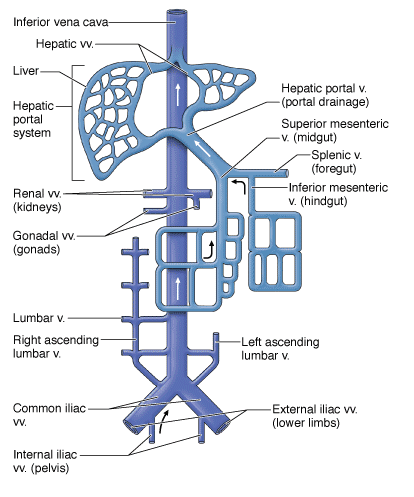
Venous blood that is returned to the heart from the anterior and posterior abdominal walls and the retroperitoneal organs via the superior or inferior vena cava.
- Inferior epigastric veins. Return blood to the heart via the inferior vena cava.
- Intercostal veins. Return blood to the heart via the superior vena cava.
- Lumbar veins. Return blood to the heart directly via the inferior vena cava or indirectly via the superior vena cava (lumbar veins may drain into the ascending lumbar veins to the azygos system of veins to the superior vena cava).
Venous blood from the gut tube and its derivatives returns to the heart via the hepatic portal vein to the liver. In other words, venous blood from the gut tube reaches the inferior vena cava after coursing through the liver.
- Foregut. Branches from the gastric and splenic veins to the portal vein.
- Midgut. Branches from the superior mesenteric vein to the portal vein.
- Hindgut. Branches from the inferior mesenteric vein to the portal vein.
The portal system drains venous blood from the distal end of the esophagus, stomach, small and large intestines, proximal portion of the rectum, pancreas, and spleen or metabolic processing before the blood returns to the heart. It is relevant for first-pass metabolism of medications and explains why GI cancers tend to metastasize to the liver. It is so vascular that it is the most common organ to have metastases from other malignancies. A malignancy found in the liver is more likely to be of metastatic than primarily hepatic origin, especially if there are multiple growths.
The portal system is the venous counterpart to areas supplied by the celiac trunk and the superior and inferior mesenteric arteries.
Blood Flow of the Portal System
The liver is unique in that it receives both nutrient-rich deoxygenated blood (portal vein) and oxygenated blood (hepatic arteries). The portal vein branches as it enters the liver, where its blood percolates around hepatocytes in tiny vascular channels known as sinusoids. Hepatocytes detoxify the blood, metabolize fats, carbohydrates, and drugs, and produce bile. The sinusoids receive deoxygenated blood from the portal veins (provide blood for metabolism and detoxification) and oxygenated blood from the hepatic arteries (provide oxygen for hepatocytes). Blood exits the sinusoids into a central vein, which empties into the hepatic veins and ultimately into the inferior vena cava, which passes through the diaphragm before entering the right atrium of the heart.
Oral drugs travel throughout the gastrointestinal tract, where they are absorbed by the small intestine. These drugs then travel to the liver via the hepatic portal system, where they are metabolized before entering the systemic circulation. Because of hepatic metabolism, the concentration of oral drugs is reduced before entering the systemic circulation. This is known as the first-pass effect. Therefore, drugs that are inactivated by the liver (e.g., nitroglycerin) must be administered by a different method. For example, nitroglycerin is administered sublingually (absorption under the tongue) because, if swallowed, the liver inactivates the drug before it can enter the systemic circulation.
Veins of the Portal System
Veins of the portal system generally mirror the arterial branches of the celiac trunk and the superior and inferior mesenteric arteries. The major veins of the portal system are as follows:
- Splenic vein. Drains blood from the foregut, including the spleen, pancreas, and part of the stomach. The splenic vein courses deep to the pancreas.
- Superior mesenteric vein. Drains blood from the midgut and part of the foregut. The superior mesenteric vein is located to the right of the superior mesenteric artery as it courses over the third part of the duodenum.
- Gastro-omental veins. Drain blood from the greater curvature of the stomach into the superior mesenteric vein.
- Inferior mesenteric vein. Drains blood from the hindgut, including the proximal third of the rectum. The inferior mesenteric vein usually drains into the superior mesenteric vein, inferior to its union with the portal vein.
- Portal vein. Collects blood from the foregut, midgut, and hindgut. The portal vein is located deep to the hepatic artery and cystic duct and is formed by the union of the superior mesenteric vein and splenic vein, deep to the neck of the pancreas.
- Gastric veins. Drain blood from the lesser curvature of the stomach into the portal vein.

A. The portal venous system. B. The three primary portal–caval anastomoses.
Portal–caval anastomoses occur at regions of the gastrointestinal tract that are drained by both the portal and systemic (-caval) systems. The principal portal–caval anastomoses are as follows:
-
Distal portion of the esophagus. The left gastric vein of the hepatic portal system drains blood from the distal portion of the esophagus. However, most of the blood drained from the esophagus is through the esophageal veins, which drain into the azygos (caval) vein.
-
Anterior abdominal wall. The paraumbilical veins drain the tissue surrounding the umbilicus: Embryologically, these veins communicated with the umbilical veins. These connections may reopen during chronic portal hypertension. Normally in the adult, most of the venous drainage is from the inferior epigastric veins.
-
Rectum. The proximal portion of the rectum is drained via the superior rectal vein, which drains into the inferior mesenteric vein of the hepatic portal system. However, the remainder of the rectum is drained by the middle rectal vein (branch of the internal iliac vein) and inferior rectal vein (branch of the internal pudendal vein).
When hepatocytes are damaged (e.g., due to disease, alcohol, or drugs), the liver cells are replaced by fibrous tissue, which impedes the flow of blood through the liver (cirrhosis). When the hepatic portal system is blocked, the return of blood from the intestines and spleen through the liver is impeded, resulting in portal hypertension. Therefore, veins that usually flow into the liver are blocked. Consequently, blood pressure in the blocked veins increases, causing them to dilate and gradually reopen previously closed connections with the caval system.
Veins in the distal portion of the esophagus begin to enlarge (esophageal varices); veins in the rectum begin to enlarge (internal hemorrhoids); and in chronic cases, the veins of the paraumbilical region enlarge (caput medusa).
Sites of Portal-Caval Venous Anastomoses1
| Portal Venous Drainage | Vena Cava Venous Drainage | Sign/Symptom | |
|---|---|---|---|
| Esophagus | Left gastric vein | Hemiazygous vein | Esophageal varices, bleeding |
| Rectum | Superior rectal vein | Inferior rectal vein | Hemorrhoids |
| Anterior abdominal wall | Paraumbilical vein | Intercostal vein | Caput medusa |
| Retroperitoneal | Duodenal, pancreatic, right and left colic veins | Lumbar vein | Intestinal bleeding |