
- Omentum
- Innervation and Vascular Supply of the Peritoneum
- Organization of the Abdominal Viscera
- Retroperitoneum
The omentum refers to modified mesenteries associated with the stomach and liver.
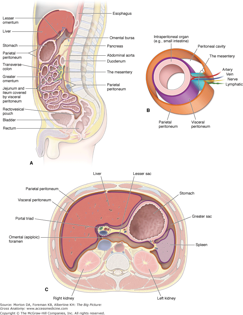
- Greater omentum. An apron-like fold of mesentery that attaches between the transverse colon to the greater curvature of the stomach.
- Lesser omentum. Mesentery that attaches between the liver, stomach, and proximal portion of the duodenum. As a result, the lesser omentum is also referred to as the hepatogastric ligament and hepatoduodenal ligament. The lesser omentum forms a sac known as the omental bursa, which forms a subdivision of the peritoneal cavity known as the lesser sac. The greater sac is the remaining part of the peritoneal cavity. The greater and lesser sacs communicate with each other through the epiploic foramen (of Winslow).
The neurovascular and lymphatic supply of the peritoneum course to and from the posterior abdominal wall and gut tube through the two-layered mesentery
(Figure B). The vascular supply to the parietal peritoneum is through the same vessels that supply the abdominal body wall, mainly the intercostal, lumbar, and epigastric vessels. The vascular supply to the visceral peritoneum is through vessels that arise from the abdominal aorta. These vessels also supply the organs in the abdominal cavity.
The nerves supplying the parietal peritoneum are the same that supply the body wall (intercostal nerves). The parietal peritoneum receives somatic sensory innervation. Somatic pain is sharp, focused, and specific. The visceral peritoneum and abdominal organs receive sensory innervation by the visceral afferents that accompany the autonomic nerves (sympathetic and parasympathetic). Visceral pain is dull, diffuse, and nonspecific.
 The parietal and visceral peritoneum are innervated by different modalities of sensory neurons; that is, parietal peritoneum via somatic innervation and visceral peritoneum via visceral innervation. Therefore, pain experienced in the parietal peritoneum is sharp, focused, and specific. In contrast, pain experienced in the visceral peritoneum is dull, diffuse, and nonspecific.
- Intraperitoneal. Viscera that are suspended from the abdominal wall by mesenteries. Intraperitoneal organs are surrounded by visceral peritoneum (e.g., stomach).
- Retroperitoneal. Viscera that are not suspended from the abdominal wall by mesenteries. Retroperitoneal organs are covered on one of their surfaces by parietal peritoneum (e.g., kidney).
Surgical procedures involving organs located in the retroperitoneal space can be accessed through the body wall, superficial to the parietal peritoneum. For example, to access organs in the retroperitoneal space, such as the kidney, a lateral incision may be made through the muscles of the body wall, leaving the parietal peritoneum intact. This approach reduces the risk of infection and peritonitis because the peritoneal cavity is not entered.
Several abdominal structures, however, are situated in the retroperitoneum (at least in part). These are the —S uprarenal glands (adrenals), A orta–inferior vena cava (IVC), D uodenum (second and third parts), P ancreas, U reters, C olon (ascending and descending), K idney, E sophagus, and R ectum.