
Vertebral Column
Lumbar Spine
https://www.shutterstock.com/search/axis+vertebrae
- Overview
- Spinal Curves
- Parts of the Spine
- Vertebral Articulations
- Vertebral Ligaments and Joints
- Study Question
The spine acts as a conduit for precious neural structures and possesses the physiological capacity to act as a crane for lifting and a crankshaft for walking.
It protects the spinal cord.
The spine is composed of the vertebral bones and intervertebral disks.
There are 7 cervical (C), 12 thoracic (T), and 5 lumbar (L) vertebrae. The sacrum is a fusion of 5 sacral (S) vertebrae, and there are small rudimentary coccygeal vertebrae.
At each vertebral level, paired spinal nerves exit the central nervous system.
The lumbar vertebrae are the 5 largest and strongest of all vertebrae in the spine. The strongest stabilizing muscles of the spine attach to the lumbar vertebrae.
Spinal Curves
The spine is made up of three segments. When viewed from the side, these segments form three natural curves. The "c-shaped" curves of the neck (cervical spine) and lower back (lumbar spine) are called lordosis. The "reverse c-shaped" curve of the chest (thoracic spine) is called kyphosis.
These curves are important to balance and they help us to stand upright. If any one of the curves becomes too large or small, it becomes difficult to stand up straight and our posture appears abnormal.
Abnormal curvatures of the spine are also referred to as spinal deformity. These types of conditions include kyphosis of the thoracic spine ("hunchback") and lordosis of the lumbar spine ("swayback").
Scoliosis is another type of spinal deformity. When viewing the spine from the front or back, scoliosis is a sideways curvature that makes the spine look more like an "S" or a "C" than a straight "I."
The anterior part of the spine consists of cylindrical vertebral bodies separated by intervertebral disks and held together by the anterior and posterior longitudinal ligaments.
The intervertebral disks are composed of a central gelatinous nucleus pulposus surrounded by a tough cartilaginous ring, the annulus fibrosis. Disks allow the bony vertebrae to move easily upon each other.
Disks allow the bony vertebrae to move easily upon each other.
Vertebral anatomy. (From A Gauthier Cornuelle, DH Gronefeld: Radiographic Anatomy Positioning. New York, McGraw-Hill, 1998; with permission.)

[Desiccation of the nucleus pulposus and degeneration of the annulus fibrosus increase with age and result in loss of disk height. ]
The disks are largest in the cervical and lumbar regions where movements of the spine are greatest. The anterior spine absorbs the shock of bodily movements such as walking and running and, with the posterior spine, protects the spinal cord and nerve roots in the spinal canal.
Sagittal section through lumbar vertebrae.
The disks are largest in the cervical and lumbar regions where movements of the spine are greatest. The anterior spine absorbs the shock of bodily movements such as walking and running and, with the posterior spine, protects the spinal cord and nerve roots in the spinal canal.
Region |
Number of Spinal Nerves |
Number of Vertebrae |
|---|---|---|
Cervical |
8 |
7 |
Thoracic |
12 |
12 |
Lumbar |
5 |
5 |
Sacral |
5 |
5 (fused) |
Coccygeal |
1 |
3–4 |
Each vertebra is referred to by the first letter of its region. For example, the fourth cervical vertebra is referred to as the C4 vertebra.
- Seven cervical vertebrae. The C1 vertebra, also called the “atlas,” articulates with the skull. The articulation between the occiput and the first cervical vertebra (the atlantooccipital joint) allows for approximately one-third of flexion and extension and one-half of lateral bending of the neck [1].
- The C2 vertebra is also called the “axis” because, through its synovial joint articulation with C1, it provides a side-to-side rotation, as in the head movement indicating “no.” A unique characteristic of cervical vertebrae is that each transverse process has its own transverse foramen for the transmission of the vertebral arteries from the subclavian artery to the brain.
- The articulation between the first and second cervical vertebrae (the atlantoaxial joint) allows for 50 percent of rotational range of motion. The articulations between the second and seventh cervical vertebrae allow for approximately two-thirds of flexion and extension, 50 percent of rotation, and 50 percent of lateral bending.
-

- Twelve thoracic vertebrae. On each side of a thoracic vertebra, there is a rib that articulates with the vertebral body and transverse process. Thus, there are 12 pairs of ribs articulating with 12 thoracic vertebrae.
- Five lumbar vertebrae. These massive vertebrae form the support to the lower part of the back.
- Five sacral vertebrae. These vertebrae are fused into one bone called the sacrum.
- Coccygeal vertebrae. There are three to four fused coccygeal vertebrae that form the coccyx, or “tailbone.”

A. Posterior view of the vertebral column. B. A typical thoracic vertebra. C. Two articulated vertebrae showing the ligaments. D. Lateral view of two vertebrae demonstrating intervertebral discs as shock absorbers. Observe how the facet joints facilitate flexion and extension of the vertebral column.
-
These vertebrae, along with their ligaments and intervertebral discs, form the flexible, protective, and supportive vertebral column that contains the spinal cord.
The anterior spine consists of cylindrical vertebral bodies separated by intervertebral disks and held together by the anterior and posterior longitudinal ligaments. The intervertebral disks are composed of a central gelatinous nucleus pulposus surrounded by a tough cartilaginous ring, the annulus fibrosis. Disks are responsible for 25% of spinal column length and allow the bony vertebrae to move easily upon each other
Vertebral anatomy. (From A Gauthier Cornuelle, DH Gronefeld: Radiographic Anatomy Positioning. New York, McGraw-Hill, 1998)
Spinal column. (From A Gauthier Cornuelle, DH Gronefeld: Radiographic Anatomy Positioning. New York, McGraw-Hill, 1998)

The disks are largest in the cervical and lumbar regions where movements of the spine are greatest. The anterior spine absorbs the shock of bodily movements such as walking and running and, with the posterior spine, protects the spinal cord and nerve roots in the spinal canal.
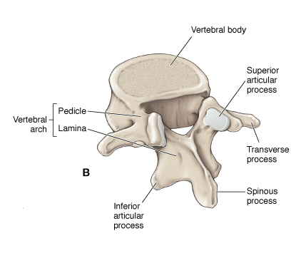
The posterior spine consists of the vertebral arches and processes. Each arch consists of paired cylindrical pedicles anteriorly and paired lamina posteriorly. The vertebral arch also gives rise to two transverse processes laterally, one spinous process posteriorly, plus two superior and two inferior articular facets. The apposition of a superior and inferior facet constitutes a facet joint. The posterior spine provides an anchor for the attachment of muscles and ligaments. The contraction of muscles attached to the spinous and transverse processes and lamina works like a system of pulleys and levers that results in flexion, extension, and lateral bending movements of the spine.
Vertebrae
The spine is made up of bones, called vertebrae, which are stacked on top of one another and create the natural curves of the back.
These bones connect to create a canal that protects the spinal cord.
The spine from the front.
The spine from the back.
The cervical spine is made up of seven small vertebrae that begin at the base of the skull and end at the upper chest. The thoracic spine is made up of 12 vertebrae that start from the upper chest to the middle back and connect to the rib cage. The lumbar vertebra consists of five larger vertebrae. These vertebrae are larger because they carry more of your body's weight.
Parts of the lumbar spine.
Spinal Cord and Nerves
The spinal cord extends from the skull to your lower back and travels through the middle part of each stacked vertebra, called the central canal. Nerves branch out from the spinal cord through openings in the vertebrae and carry messages between the brain and muscles.
The spinal cord ends around the first and second lumbar vertebrae in the lower back and continues as nerve roots. This bundle of nerve roots is called the cauda equina. They exit the spinal canal through openings in the vertebrae (foramen), just like other nerve roots. In the pelvis, some of the nerves group into the sciatic nerve, which extends down the leg.
The cauda equina.
Muscles and Ligaments
These provide support and stability for your spine and upper body. Strong ligaments connect your vertebrae and help keep the spinal column in position.
Intervertebral Disks
Intervertebral disks sit in between the vertebrae. They are flat and round, and about a half inch thick.
Intervertebral disks are made up of two components:
- Nucleus pulposus. The nucleus pulposus is jelly-like and makes up the center of the disk. The jelly is partly made of water and gives the disk flexibility and strength.
- Annulus fibrosus. This is the flexible outer ring of the disk. It is made up of several layers, similar to elastic bands.
When you are standing or moving, weight is put onto the nucleus. In response, the nucleus expands. The annulus holds the nucleus in place. This allows movement to take place, yet maintains the strength of the spine. In effect, disks act as shock absorbers for the spine.
The intervertebral disk is a very important structure. Many nerve endings supply the annulus and, as a result, an injured annulus can cause pain.
Healthy intervertebral disk (cross-section view).
Facet Joints
Between the back of the vertebrae are small joints that also help your spine move. These facet joints have a cartilage surface, very much like a hip or a knee joint does. The facet joints are important for allowing rotation of the spine but may develop arthritis and become a source for low back or neck pain.
+++++++++++++++
- Body. The anterior vertebral region that is the primary weight-bearing component of the vertebrae.
- Vertebral arch. Arches posteriorly from the vertebral body and forms the vertebral foreman, which contains the spinal cord.
- Pedicle. Part of the vertebral arch that joins the vertebral body to the transverse process.
- Transverse processes. Extend laterally from the junction of the pedicle and lamina.
- Superior and inferior articular facets. Form synovial zygapophyseal (facet) joints with the vertebrae above and below.
- Laminae. The paired posterior segments of the vertebral arch that connect the transverse processes to the spinous process.
- Spinous processes. The posteriorly projecting tip of the vertebral arch. These processes are easily palpated beneath the skin.
- Vertebral foramen. The space in which the spinal cord, its coverings, and the rootlets of the spinal nerves lie. The series of vertebral foramina form the vertebral canal.
- Intervertebral foramina. Bilateral foramina that form the space between pedicles of adjacent vertebrae for passage of spinal nerves.
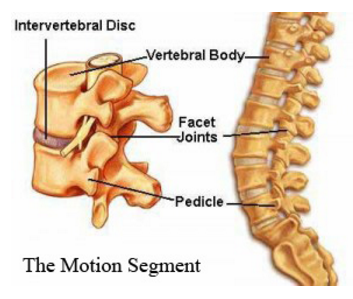 There are two types of articulations that enable the 33 vertebrae to not only provide support, protection, and absorb shock, but also to provide flexibility.
These two articulations are the intervertebral discs and the facet joints.
- Intervertebral discs. Located between vertebrae, from the second cervical disc down to the first sacral vertebra (Figure 1-1D). The discs permit a limited amount of movement between adjacent vertebrae. Intervertebral discs are composed of an annulus fibrosus and a nucleus pulposus. The anulus fibrosus is the tough hyaline cartilaginous rim, whereas the nucleus pulposus is the softer fibrocartilaginous core. The disc can bear weight because spread of the nucleus pulposus is constrained by the anulus fibrosus. The nucleus is composed of proteoglycan megamolecules that can imbibe water to a capacity approximately 250% of their weight.
 Damage to the anulus fibrosus may allow the softer nucleus pulposus to bulge or herniate, which may compress the segmental nerve roots. This is often referred to as a “slipped disc.” The symptoms of this condition will depend upon the level at which the rupture occurs and the structures that are affected. For example, compression of the L4 nerve may cause weakness in ankle dorsiflexion due to the L4 innervation of the anterior tibialis muscle in the leg, or lack of cutaneous sensation in the skin over the knee due to the L4 dermatome.
- Superior and inferior articular facets of adjacent vertebrae. These joints provide varying amounts of flexibility.
A disc and its associated facet joints make up an individual motion segment. In between the disc and the facet joints lies the spinal cord or nerves and to the sides are bony struts called pedicles (see image below). Sprouting off from the spinal cord and shooting out from underneath the pedicles are the nerves, which in the lumbar spine mostly form the femoral, and sciatic nerves and in the cervical spine form the brachial plexus (the nerves which travel down the arms). In the thoracic spine the nerves supply sensation to bands of skin radiating around to the front of the chest wall and abdomen, and move the intercostal muscles to that we can inflate our lungs.
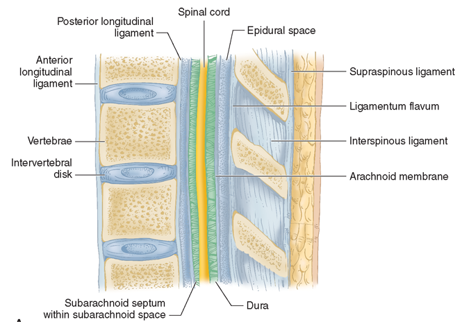
The vertebral column is stabilized by ligaments that run between vertebrae.
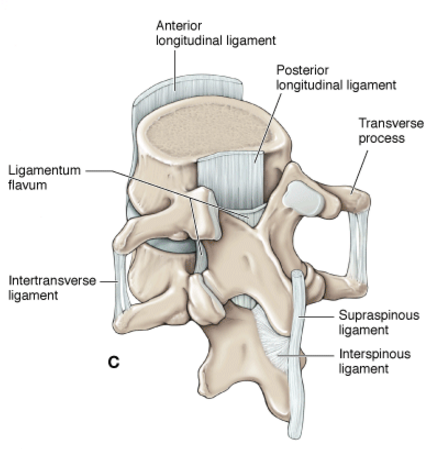
Some of the ligaments that limit vertebral flexion (bending forward) include the following:
- Ligamentum flavum. Connects paired laminae of adjacent vertebrae.
- Supraspinous ligament. Connects the apices of the spinous processes from C7 to the sacrum.
- Interspinous ligament. Connects adjoining spinous processes.
- Nuchal ligament. Extends from the external occipital protuberance along the spinous processes of C1–C7.
- Posterior longitudinal ligament. Courses longitudinally, down the posterior surface of the vertebral bodies within the vertebral canal. It supports the intervertebral disc posteriorly, thus reducing the incidence of herniations that may compress the spinal cord and cauda equina.
- Anterior longitudinal ligament. Courses longitudinally along the anterior surface of the vertebral bodies limiting vertebral extension.
Which of the following paired muscles of the back is primarily responsible for extension of the vertebral column?
Answer (Click)
Content 2
Content 3