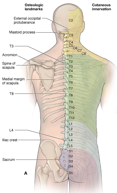
The skin of the back is thick, with increasing thickness toward the nape of the neck.
The skin of the back is segmentally innervated by cutaneous nerves that originate from the dorsal rami. Dorsal rami contain both motor and sensory neurons as they branch from each level of the spinal cord and course posteriorly in the trunk.
|
A. Surface anatomy of the back showing bony landmarks on the left and cutaneous nerves on the right. B. Axial section of the back showing the dorsal rami transmitting sensory neurons from the skin of the back to the spinal cord. Normal curvatures of the vertebral column in a newborn (C) and in an adult (D). |
The following structures are easily palpated through the skin.
 (Figure 1-1A):
- Cervical vertebrae. The first prominent spinous process that is palpable is C7 (vertebra prominens). Cervical spines 1 to 6 are covered by the ligamentum nuchae, a large ligament that courses down the back of the neck and connects the skull to the spinous processes of the cervical vertebrae.
- Thoracic vertebrae. The most prominent spine is the T1 vertebra; other vertebrae can be easily recognized when the trunk is flexed anteriorly. Thoracic vertebrae have long spines that point downward so that each spinous process is level with the body of the inferior vertebra. The spines can be counted downward from C7 and T1, or from a line joining the iliac crests at the L4 vertebral level and then counting upward from that site.
- Lumbar vertebrae. The body of L3 is approximately at the center of the body from superior to inferior and medial to lateral. When a person is in an upright posture, most vertebral spines are not obvious because they are covered by the erector spinae muscles in the cervical and lumbar regions.
- Sacrum. The spines of the sacrum are fused together in the midline to form the median sacral crest. The crest can be felt beneath the skin in the uppermost part of the gluteal cleft between the buttocks. The sacral hiatus is situated on the posteroinferior aspect of the sacrum and is the location where the extradural space terminates. The hiatus lies approximately 5 cm above the tip of the coccyx and beneath the skin of the natal cleft.
- Coccyx. The tip of the coccyx is located in the upper part of the natal cleft and can be palpated approximately 2.5 cm posterior to the anus. The anterior surface of the coccyx can be palpated via the anal canal.
- Ilium. The crest of the ilium is located at the L4 vertebral level and is easily palpated. Regardless of the size or weight of a person, the posterior iliac spines can be palpated because they lie beneath a skin dimple at the S2 vertebral level and the middle of the sacroiliac joint.
- Scapulae. The scapulae overlie the posterior portion of the thoracic wall, covering the upper seven ribs. The superior angle can be palpated at the T1 vertebral level, the spine of the scapula at T3, and the inferior angel at T7. The acromion forms the lateral end of the scapular spine and is easily palpated.
|

 .
.