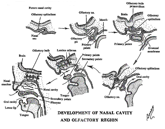
The placode invaginates to form the olfactory pit, which later gives rise to the olfactory epithelium.
Neural crest cells contribute supporting cells and some mesenchymal elements.

The primordia of the olfactory system consist of 2 placodes, a thickening of ectoderm, situated above the stomodeum and below and lateral to the forebrain.
The primordia appear about day 30, after those of the optic and otic placodes.
At this time, too, the neural tube is completely close
The placode invaginates to form the olfactory pit, which later gives rise to the olfactory epithelium.
Neural crest cells contribute supporting cells and some mesenchymal elements.
The olfactory pit deepens and becomes continuous with the primitive nasal cavity.
The lining epithelium differentiates into:
Olfactory receptor neurons (ORNs) – bipolar neurons with apical cilia that detect odorants.
Supporting (sustentacular) cells – provide metabolic and structural support.
Basal cells – stem/progenitor cells that continually regenerate olfactory receptor neurons (unique for a sensory system).
ORN axons extend through the cribriform plate and form the olfactory nerve (CN I).
These axons induce the formation of the olfactory bulb from the rostral telencephalon.
In the bulb, axons synapse in glomeruli with mitral and tufted cells (secondary neurons of the olfactory pathway).
This target-dependent maturation means the bulb and ORNs co-develop: ORN axons need the bulb for guidance, and the bulb requires ORN innervation for proper organization.
The placodal cells of the olfactory epithelium differentiate into neurosensory cells and eventually give origin to olfactory nerve fibers
Mitral and tufted cell axons form the olfactory tract, projecting to:
Piriform cortex
Amygdala
Entorhinal cortex
Orbitofrontal cortex (indirectly via thalamus)
Near the end pf month 3, the mesenchyme between the sensory epithelium and the bulb gives rise to a cartilaginous structure, the lamina cribrosa of the ethmoid bone which is eventually organized around the olfactory nerve networks and separates them into a number of bundles. The cartilage ossifies here to form the cribriform plate of the ethmoid through which the nerves pass to enter the olfactory bulbs
The olfactory bulb elongates, and eventually the extension of the ventricular cavity into it becomes obliterated
Cells in the bulb, around which the olfactory nerve fibers terminate and synapse, give origin to secondary olfactory fibers which grow centrally and form the olfactory tract
The olfactory tract terminates in the region of the piriform cortex
Fibers of the olfactory nerves are entirely of placodal origin and their cell bodies remain in the olfactory epithelium.1
Olfactory receptor neurons are continually replaced throughout life (every 30–60 days).
This regenerative capacity is sustained by basal stem cells in the olfactory epithelium.
The system remains plastic, allowing adaptation to new odorants and recovery after injury.
Kallmann syndrome – failure of olfactory axons and GnRH neurons to migrate properly → anosmia + hypogonadotropic hypogonadism.
Congenital anosmia – isolated absence of olfactory bulb/tract.
Neurodegenerative diseases (e.g., Parkinson’s, Alzheimer’s) – early olfactory deficits due to degeneration of olfactory structures.
++++++++++++++
